Chemistry of Blood Groups


Chemistry of Blood Groups


types of blood groups


Abstract

International Society of Blood Transfusion has recently recognized 33 blood group systems. Apart from ABO and Rhesus system, many other types of antigens have been noticed on the red cell membranes. Blood grouping and cross-matching is one of the few important tests that the anesthesiologist orders during the perioperative period. Hence, a proper understanding of the blood group system, their clinical significance, typing and cross-matching tests, and current perspective are of paramount importance to prevent transfusion-related complications It is firmly established that ABO and rhesus (Rh) genes and phenotypes remain the most significant blood factor in clinical applications, it also proves to be a valuable assets for determining human migration patterns and origins. To determine the prevalence of ABO and Rh blood groups based on the antigenic presence on the surface of red blood cells concerning gender and calculate the allele frequency of the blood groups. Globally, approximately 700 type red cell antigens have been identified till now. ABO and Rh blood groups play an important role in the process of blood transfusion, resolving certain medico-legal issues, parental testing, and various genetic studies.

Introduction

Human blood Cells:Normally, 7-8% of human body weight is from blood.  In adults, this amounts to 4.5-6 quarts of blood.  This essential fluid carries out the critical functions of transporting oxygen and nutrients to our cells and getting rid of carbon dioxide, ammonia, and other waste products.  Also, it playsa role in our immune system and in maintaining a relatively constant body temperature. Blood is a highly specialized tissue composed of more than 4,000 different kinds of components.  Four of the most important ones are red cells, white cells, platelets, and plasma. Red cells or Human erythrocytes or "red cells" (cell width around .0003 inches) Red cells, or erythrocytes click this symbol to hear the first term articulated, are moderately enormous infinitesimal cells without cores. In this last attribute, they are like the crude prokaryotic cells of microbes. Red cells ordinarily make up 40-half of the all out blood volume. They transport oxygen from the lungs to the entirety of the living tissues of the body and divert carbon dioxide. The red cells are delivered consistently in our bone marrow from undifferentiated organisms at a pace of around 2-3 million cells each second. Hemoglobin click this symbol to hear the first term articulated is the gas moving protein atom that makes up 95% of a red cell. Every red cell has around 270,000,000 iron-rich hemoglobin particles. Individuals who are pale by and large have an inadequacy in red cells, and consequently feel exhausted because of a lack of oxygen.White CellsWhite cells: 
White cells, or leukocytes click this symbol to hear the first term articulated, exist in factor numbers and types yet make up a tiny piece of blood's volume- - regularly just about 1% in solid individuals. Leukocytes are not restricted to blood. They happen somewhere else in the body also, most prominently in the spleen, liver, and lymph organs. Most are created in our bone marrow from the very sort of immature microorganisms that produce red platelets. Others are delivered in the thymus organ, which is at the base of the neck. Some white cells (called lymphocytes click this symbol to hear the former term articulated) are the people on call for our invulnerable system.Blood is comprised of different parts, including red platelets, white platelets, platelets, and plasma .

Platelets:

Platelets, or thrombocytes, are cell fragments without nuclei that work with blood clotting chemicals at the site of wounds.  They do this by adhering to the walls of blood vessels, thereby plugging the rupture in the vascular wall.  They also can release coagulating chemicals that cause clots to form in the blood that can plug up xiinarrowed blood vessels.
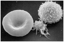
erythrocyte (left), thrombocyte
(center), and leukocyte (right) 

Plasma:

is the moderately clear, yellow-colored water (92+%), sugar, fat, protein, and salt arrangement which conveys the red cells, white cells, and platelets. Regularly, 55% of our blood's volume is comprised of plasma. As the heart siphons blood to cells all through the body, plasma carries sustenance to them and eliminates the byproducts of digestion. Plasma additionally contains blood coagulating factors, sugars, lipids, vitamins,minerals, chemicals, compounds, antibodies, and different proteins 

Introduction to Human Factors Blood Groups: 

The most notable and medicinally significant blood classifications are in the ABO gathering. They were found in 1900 and 1901 at the University of Vienna by Karl Landsteiner during the time spent attempting to realize why blood bondings here and there cause demise and at different occasions save a patient.(Schwarz and Dorner, 2003) 

The ABO Blood bunch System: 

All people and numerous different primates can be composed for the ABO blood gathering. There are four chief sorts: A, B, AB, and O. there are two antigens and two antibodies that are generally answerable for the ABO types. The particular blend of these four segments decides a person's sort much of the time. The table beneath shows the potential changes of antigens and antibodies with the comparing ABO type ("yes" demonstrates the presence of a part and "no" demonstrates its nonappearance in the blood of a person).


For instance, individuals with type A blood will have the An antigen on the outside of their red cells accordingly, hostile to An antibodies won't be created by them since they would annihilate their blood. Notwithstanding, if B type blood is infused into their frameworks, against B antibodies in their plasma blood of outsider protein.

Fig.2.ABO Blood group

It is simple and reasonable to decide a person's ABO type from a couple of drops of blood. A serum containing hostile to An antibodies is blended in with a portion of the blood. Another serum with hostile to B antibodies is blended in with the excess example. Regardless of whether agglutination happens in either test shows the ABO type. It is a straightforward interaction of end of the prospects. For example, if a person's blood test is agglutinated by the counter An immune response, yet not the counter B neutralizer, it implies that the An antigen is available however not the B antigen. Hence, the blood classification is A. 

 Genetic Inheritance Patterns: 

Exploration completed in Heidelberg, Germany by Ludwik Hirszfeld and Emil von Dungeon in 1910 and 1911 demonstrated that the ABO blood classifications are acquired. We currently realize that they are dictated by qualities on chromosome 9, and they don't change because of natural impacts during life. A person's ABO type results from the legacy of 1 of 3 alleles (A, B, or O) from each parent. The potential results are appeared beneath.

Parent Alleles
       
graphic arow pointing to the possible parental alleles for the ABO blood system along the top row and the left column

A

B

O

A

AA
(A)

AB
(AB)

AO
(A)

B

AB
(AB)

BB
(B)

BO
(B)

O

AO
(A)

BO
(B)

OO
(O)

The possible ABO alleles for one
parent are in the top row and the
alleles of the other are in the left
column.  Offspring genotypes
are shown in black.  Phenotypes
are red.

GENES ENCODING GLYCOSYLTRANSFERASES INVOLVED IN SYNTHESIS OF ABO BLOOD GROUP CARBOHYDRATE ANTIGEN STRUCTURES IN HUMANS.

The single ABO quality locus in people has three allele's sorts A, B, and O. Loci An and B encode two distinctive dynamic glycosyltransferases that catalyze the biosynthesis of An and B terminal sugar antigen groupings, utilizing O antigen carb structure as an antecedent. The O locus is encoding nonfunctional catalysts. Both An and B chemicals have a place with the glycosyltransferase family numbered as 6, as per the HUGO Gene Nomenclature Committee which is the solitary overall position that allocates normalized terminology to human qualities. The human ABO quality is situated on chromosome 9 position. The cytogenetic band is named. The genomic organizes are (GRCh38). This quality has a place with the glycosyltransferase family 6. The ABO quality has 7 exons with exon 7 having the biggest coding grouping. (Seto et al., 1995)The coding succession has more than 18-kilo base pairsand exons size range beA B C. ABO blood bunch antigen glycan groupings. ABO blood bunch antigen glycan arrangements are introduced in shortened substance letter structure, synthetic primary structure, and schematic structure as per, Gal: galactose; Fuc: fucose; GalNAc: N-acetyl galactosamine.ABO quality construction. Schematic introduction of ABO quality chromosomal area and exons positions encoding glycosyltransferase An and B. 

Historical Background: 

It was not until the year 1900, when Karl Landsteiner at the University of Vienna, found why some blood bondings were fruitful while others could be dangerous. Landsteiner found the ABO blood bunch framework by blending the red cells and serum of every one of his staff. He showed that the serum of certain individuals agglutinated the red cells of others. From these early examinations, he recognized three sorts, called A, B and (C was later to be re-named O for the German "Ohne", signifying "without", or "Zero", "invalid" in English). The fourth less regular blood bunch AB was found a year later. In 1930, Landsteiner got the Nobel Prize in physiology and medication for his work.(Seto et al., 1995)The quality that decides the human ABO blood classification is situated on chromosome 9 and is called ABO glycosyltransferase. The ABO locus has three primary allelic structures: A, B, and O, as referenced above and every one of them is answerable for the creation of its glycoprotein. As examinations have exhibited on monkeys, human blood bunches are exceptionally old hereditary pointers that have developed more than a few million years. In view of the essential races theory, it was believed that in the three significant races of man, blood bunches An in Europe, B in Asian, lastly O in South America have been arisen and progressively because of the movement and blending of the races, turned into the current circumstance. However, we realize that in every landmass, the confined populaces are seen that have totally extraordinary blood gatherings. For instance, there is a generally high pervasiveness of blood bunch O in Siberian occupants; likewise this blood bunch is basic in certain regions. 

Percentage of blood groups in monkeys (collected by Kramps 1960)

As indicated by another theory, the rise of all blood bunches An and B and their subgroups, are come about because of progressive transformations, from a fundamental and regular blood gathering, which is the O gathering, and have been spread more than a long period of time. 

In view of this hypothesis, the old races have O blood gatherings, for example, Red Indians of South America, and Eskimos that among them the recurrence of O blood bunch is between 75–100%. While in the vast majority of ongoing ethnic gatherings An and B blood bunches are dominant.In another speculation, the primary blood bunch had been the AB blood gathering, which progressively and after some time because of hereditary changes was brought about An and B lastly O blood gatherings. Base on this hypothesis, maybe a couple million years back all individuals have had type O blood just, which is more safe against numerous irresistible diseases.Based on the subsequent theory, the principal blood bunch had been AB, which continuously has been brought about An and B and O blood gatherings. 

After the revelation of the main human blood gatherings (ABO) by Karl Landsteiner in 1901, progressively from 1927, other blood bunches were likewise found and detailed in which its assortment is given. Mention that Landsteiner along with his American associate Alexander Wiener found the Rh blood gathering and announced it in 1940, 1941.
blood cells

What are the symptoms of isoimmunization?

The mother won't have manifestations from isoimmunization however for the infant indications can go from gentle to perilous. Indeed, even mellow, the contrariness obliterates the red platelets without demonstrating different impacts. At the point when the cycle is sufficiently extreme, the child can turn out to be exceptionally pale and, now and again may pass on. After birth, the child's skin and whites of the eyes will seem yellow (jaundice) and the infant will have low muscle tone (hypotonia) and torpidity.

How is isoimmunization diagnosed?

Ladies in danger for isoimmunization can be distinguished at pre-birth visits with tests that measure blood classification, Rh type, and immune response screening. Periodically the particular contradiction is analyzed before birth through amniocentesis. On the off chance that isoimmunization is analyzed, we screen the seriousness of the infant's sickliness using ultrasound. After birth, there may likewise be a positive perusing on a blood test called Coombs, higher-than-ordinary degrees of bilirubin from blood tests from the child's umbilical line, and indications of red platelet obliteration in the baby's blood.

Fig.4. the Genetic Basis of Blood Grouping in Human Population.

When recognized as having Rh-negative blood classification, the pregnant or potentially recently conveyed mother can be given RhoGAM (Rh-resistant globulin).
What is the treatment for isoimmunization?


In people, the blood bunches are constrained by a quality called quality 'I'. It has three alleles, in particular IA, IB, and i.Human blood bunch legacy is the case of codominance and various alleles.
Glycoconjugate is the overall characterization for sugars covalently connected with other synthetic species, for example, proteins, peptides, lipids andsaccharides.Glycoconjugates are shaped in cycles named glycosylation.(Daniels, 2005)Glycoconjugates are vital mixtures in science and comprise of various classes, for example, glycoproteins, glycopeptides, peptidoglycans, glycolipids, glycosides, and lipopolysaccharides. They are engaged with cell-cell communications, including cell-cell acknowledgment; in cell-lattice associations; in detoxification processes.(Robyt, 2012) 

By and large the carb part(s) assume a basic part in the capacity of a glycoconjugate; unmistakable instances of this are NCAM and bokimuj lood proteins where fine subtleties in the sugar structuredetermine cell restricting or not or lifetime in circulation.Although the significant atomic species DNA, RNA, ATP, cAMP, cGMP, NADH, NADPH, and coenzyme An all contain a carb part; by and large they are not considered as glycoconjugates.(Hult et al., 2005)Glycoconjugateisa covalent connecting of carbs antigens to protein frameworks to accomplish a drawn out immunological reaction in the body. Inoculation with glycoconjugates effectively prompted long haul safe memory against sugars antigens. Glycoconjugate antibodies presented since the 1990s haveyielded compelling outcomes against flu and meningococcus.

Glycoproteins:

Film bound glycoproteins partake in a wide scope of cell marvels, including cell acknowledgment, cell surface antigenicity, and so forth In the glycoproteins, most of the particle comprise of proteins; they have at least one oligosaccharides connected to a protein, and they generally are fanned and don't have sequential rehashes, so they are wealthy in data, shaping profoundly explicit destinations for acknowledgment and high-liking restricting by other proteins. 

Glycolipids: 

Glycolipids are film lipids in which the hydrophilic head bunches are oligosaccharides.O-N-acetylgalactosamine (O-GalNAc)The expansion of N-acetylgalactosamine (GalNAc) to a serine or threonine happens in the Golgi mechanical assembly, after the protein has been folded.The measure is performed by chemicals known as GalNAc transferases (GALNTs), of which there are 20 distinctive types.The starting O-GalNAc design can be altered by the expansion of different sugars, or different mixtures, for example, methyl and acetyl groups.(Roberts, 1988) Thrombospondin ties to the secluded glycolipid, and the glycolipid and an immunizer to this construction repress cell spreading on thrombospondin substrates. Consequently, the presence of glycoconjugates with terminal nonreducing glucuronosyl 3-sulfate associates with melanoma cell spreading on thrombospondin, while articulation of heparan sulfate proteoglycans that tight spot thrombospondin doesn't. (Miura et al., 1999) The saccharides that are connected to the polar head bunches outwardly of the cell are the ligand segments of glycolipids, and are moreover polar, permitting them to be dissolvable in the fluid climate encompassing the cell.

Fig.5.O-GalNAc core structures; Core 1, Core 2, and poly-N-acetyllactosamine structures.


The biosynthesis of N-linked glycan’s occurs via 3 major steps:

1.                  Synthesis of dolichol-linked precursor oligosaccharide

2.                  En bloc transfer of precursor oligosaccharide to protein

3.                  Processing of the oligosaccharide


Fig.6.Biosynthesis of N-Linked glycoprotein:

 The synthesis of glycan starts in the endoplasmic reticulum, continues in the Golgi, and ends at the plasma membrane, where the N-linked glycoproteins are either secreted or becomes embedded in the plasma membrane.

Biosynthesis of glycosphingolipids:

Glycosphingolipids biosynthesis starts with the blend of ceramide on the cytosolic handout of the ER. The antecedent for all cell ceramide is dihydrosphingosine whose blend is started by the buildup of L-serine and palmitoyl-CoA. The buildup response is catalyzed by L-serine palmitoyltransferase, a compound of the ER layer. The subsequent 3-ketosphinganine is decreased to dihydrosphingosine, a response catalyzed by a reductase. Dihydrosphingosine is then quickly N-acylated by an acyltransferase bringing about the arrangement of ceramide. These responses happen on the cytosolic handout of the ER layer. The glycosylation of ceramideresults in the development of glycosphingolipids of tissue cells; in the phylogenetic transformative tree erythrocytes are the latest cells to secure these antigens, since just people and arthropod gorillas express ABH antigens on their red cells. ABH or ABH-like determinants have additionally been distinguished in plants and microorganisms, yet these determinants are not under discussionheresubstances with serological specificities indistinguishable or firmly identified .
test to find blood group

O-GalNAc center constructions; Addition of N-acetylgalactosamine (GalNAc) to a serine or threonine happens in the Golgi mechanical assembly, after the protein has been collapsed. The interaction is performed by proteins known as GalNAc transferases (GALNTs), of which there are 20 unique sorts. The underlying O-GalNAc construction can be adjusted by the expansion of different sugars, or different mixtures, for example, methyl and acetyl gatherings. These alterations produce 8 center constructions known to date. Various cells have various catalysts that can add further sugars, known as glycosyltransferases, and constructions consequently change from cell to cell.Once this underlying sugar has been added, other glycosyltransferases can catalyze the option of added sugars. Two of the most widely recognized constructions framed are Core 1 and Core 2. Center 1 is framed by the expansion of a galactose sugar onto the underlying GalNAc. Center 2 comprises of a Core 1 design with an extra N-acetyl glucosamine (GlcNAc) sugar. A poly-N-acetyllactosamine construction can be shaped by the exchanging expansion of GlcNAc and galactose sugars onto the GalNAc sugar.A sugar-containing galactose and GlcNAc is known as lactose, giving the design its name.Terminal sugars on O-glycan are significant in acknowledgment by lectins and assume a vital part in the invulnerable framework. The expansion of fucose sugars by fucosyltransferases structures Lewis epitopes and the framework for blood bunch determinants. The option of a fucose alone makes the H-antigen, present in individuals with blood classification O.By adding a galactose onto this construction, the B-antigen of blood bunch B is made. (HÅRD et al., 1992)Alternatively, adding a GalNAc sugar will make the A-antigen for blood bunch ABiological elements of atomic proteins are controlled by post-translational alterations (PTMs) that tweak quality articulation and cell physiology. Notwithstanding, the part of O-connected glycosylation (O-GalNAc) as a PTM of atomic proteins in the human cell has not been recently detailed. Here, we inspected in detail the inception of O-GalNAc glycan biosynthesis, addressing a novel PTM of atomic proteins in the core of human cells, with an accentuation on HeLa cells. Utilizing solvent atomic divisions from cleaned cores, enzymatic measures, fluorescence microscopy, partiality chromatography, MS, and FRET investigations, we recognized all elements needed for the biosynthesis of O-GalNAc glycans in cores: the contributor substrate (UDP-GalNAc), atomic polypeptide GalNAc - transferases action, and a GalNAc transferase.

humanoid primates to communicate ABH antigens on their red cells. ABH or ABH-like determinants have likewise been recognized in plants and microorganisms, yet these determinants are not being talked about here. 

 ABO(H) System: 

The characters characterized in the ABO(H) framework are dependent upon severe hereditary control and accordingly are acquired as basic Mendelian prevailing characters. Event and conveyance of ABH specificities are constrained by three autonomous however firmly interrelated quality systems.The ABO Gene The three alleles A B, and 0 of this quality locus represent the four fundamental blood gatherings, A, B, AB, and O. The alleles An and B control the arrangement of An and B explicitness on erythrocytes and in emissions, though the dormant 0 allele doesn't offer ascent to any blood bunch character. The connection between ABO genotype and noticed erythrocyte aggregate is introduced in.The dominant part of An and B subgroups characterized to date are constrained by variations of An and Bgenes. The ABO locus has been restricted on chromosome 9, position. The Hh Gene The blood bunch H character is found on erythrocytes and in discharges of blood bunch 0 people and has subsequently been viewed as the result of the 0 gene.(Schenkel-Brunner, 2000) H determinants, nonetheless, are additionally present, however not in amount, in A, B, and AB subjects. Since people of genotypes M BB and AB come up short on the 0 qualities, Watkins and Morgan inferred that this particularity should be the result of a free quality, which they assigned HH substance addresses the forerunner which, affected by qualities An and B, is changed over into blood bunch An or B substance, separately. Since the latent 0 allele not the slightest bit modifies H substance, the presence of unaltered H determinants is trademark for 0 people. There is besides a proportional relationship.


1.                  ABH Antigens on Red Cells and in Plasma:

humanoid primates to communicate ABH antigens on their red cells. ABH or ABH-like determinants have likewise been recognized in plants and microorganisms, however these determinants are not being talked about here. 

 ABO(H) System: 

The characters characterized in the ABO(H) framework are dependent upon exacting hereditary control and accordingly are acquired as straightforward Mendelian predominant characters. OccurrencA, B, and H immunodominant sugars are added to forerunner chains called type 1 and type 2 chains. These chains contrast in the linkage of a galactose to a N-acetyl-D-glucosamine buildup: in sort 1 chains the linkage is beta 1 - > 3, and in sort 2 chains it is beta 1 - > 4.Research has indicated that the ABH antigens orchestrated by the red cells are just continued sort 2 binds and are joined to both glycolipids and glycoproteins. Be that as it may, some ABH antigens on red cells are obtained from the plasma (and in this way are subject to secretor qualities just as on ABH qualities). The ABH antigens obtained from the plasma are on sort 1 chains and are (glycolipids can be embedded into the layer of red cells). The site of amalgamation of plasma ABH antigens is unknown.(Watkins, 1995) 
blood group experiments

ABH Secretion: 

The term secretor, as utilized in blood banking, alludes to the discharge of ABH antigens in liquids, for example, salivation, sweat, tears, semen, and serum. In the event that individuals are secretors, they will emit antigens as per their blood gatherings. For instance, bunch O individuals will emit H antigen, bunch A people will discharge An and H antigens, and so forth Solvent 
e and dispersion of ABH specificities are constrained by three free however firmly interrelated quality systems.The ABO Gene The three alleles A B, and 0 of this quality locus represent the four principle blood gatherings, A, B, AB, and O. The alleles An and B control the arrangement of An and B explicitness on erythrocytes and in discharges, while the idle 0 allele doesn't offer ascent to any blood bunch character. The connection between ABO genotype and noticed erythrocyte aggregate is introduced in.The dominant part of An and B subgroups characterized to date are constrained by variations of An and Bgenes. The ABO locus has been confined on chromosome 9, position. The Hh Gene The blood bunch H character is found on erythrocytes and in discharges of blood bunch 0 people and has consequently been viewed as the result of the 0 gene.(Schenkel-Brunner, 2000) H determinants, in any case, are additionally present, however not in amount, in A, B, and AB subjects. Since people of genotypes M BB and AB do not have the 0 qualities, Watkins and Morgan presumed that this particularity should be the result of a free quality, which they assigned HH substance addresses the forerunner which, affected by qualities An and B, is changed over into blood bunch An or B substance, individually. Since the inert 0 allele not the slightest bit adjusts H substance, the presence of unaltered H determinants is trademark for 0 people. There is besides a proportional relationship.

1.                  Test for Secretor Status (Inhibition Test)

To test for secretor status, a restraint or balance test is finished utilizing spit. The rule of the test is that if ABH antigens are available in a solvent structure in a liquid (e.g., spit) they will kill their comparing antibodies and the antibodies can not, at this point agglutinate red cells having similar antigens. (Note: Secretor status testing is only from time to time, if at any time, done today because of the approach of more strong monoclonal composing reagents and DNA technology.)(Raza et al., 1991) 

5.2Antisera& Lectins: Antisera are utilized to figure out which antigens are on the red cells. The test utilizing antisera and the patient's red cells is known as the front cell gathering. The antisera utilized in the front gathering are acquired from plasmapheresis of benefactors invigorated with solvent antigens (A substance from pig mucosa and B substance from horse mucosa). On the other hand, monoclonal antisera can be set up from refined cell lines.

1.                 anti-A (from group B people)

The government regulations specify that anti-A be colored blue as a quality control (QC) measure.

2.                  anti-B (from group A people)

The government regulations specify that anti-B be colored yellow as a QC measure.

3.                  anti-A,B (from group O people)

Against A,B is lackluster (no color is added). Utilization of against A,B shifts from lab to lab. A few labs use it for all ABO gathering, and some utilization it to affirm bunch O composing. Other blood donation centers use it specifically as follows:to bunch babies (whose converse gathering is inconsistent and can't be utilized as a mind the front cell group).whenever errors happen between the front and opposite gathering, particularly disparities because of powerless or missing antigens. Note: monoclonal enemy of A,B is greater at identifying powerless subgroups than is human enemy of A,B.when exploring suspected hemolytic bonding responses

Main uses of anti-A,B:

1.                  To confirm the front cell group in newborns.

2.                  To help resolve ABO discrepancies.

Monoclonal Reagents: murine (mouse) monoclonal anti-A and anti-B reagents prepared using hybridoma technology are available as alternatives to human antisera. These monoclonal antisera are very specific and very potent (sensitive). They are useful when investigating both weak and extra antigens, e.g., weak subgroups of A or acquired B phenomenon.

ABH Substances of erythrocytes:

The commitment of blood-bunch dynamic glycolipids and glycoproteins to the blood-bunch ABHcharacter of human erythrocytes was explored. For that reason the blood-bunch H locales of human O cells were changed over in vitro into bunch A destinations by move of alpha-N-acetyl-D-galactosamine deposits with the guide of the blood-bunch A quality ward alpha-N-acetylgalactosaminyl transferase arranged from human A1 plasma. Upon the segment of the red cell films among water and natural dissolvable, about 5% of the name was found in the natural stage and about 20% in the water stage, in this way mirroring the appropriation of blood-bunch antigenic locales between glycosphingolipids with short carb chains and polyglycosylceramides, individually. The way that about 70% of the radioactivity remained firmly bound to the layers and must be delivered by treatment with pronase gave great proof that the greater part of blood-bunch H determinants is bound to glycoprotein material. Following these outcomes it would thus be able to be accepted that blood-bunch ABH action of human erythrocytes is resolved specially by bunch explicit glycoproteins instead of glycolipids.

Secreted Blood group substances  (Mucins):

For the two sorts of ulcers, the assessed hazard among non-secretors was comparable for blood bunch O and the other blood gatherings and the outcomes propose that the particular dangers related with blood bunch O and with non-emission duplicate each other. It is presumed that the systems by which blood bunch O and non-discharge influence the danger of building up a gastric or duodenal ulcer are identified with each other; however that they don't rely upon the presence of the blood bunch substances in the secretions.(Gibbons, 1959).

Distribution and examination of secretor status pervasiveness in people ABH antigens are available on platelets from people of the relating red cell aggregate, yet the degree to which these antigens are inborn or adsorbed stays unclear. To assess platelets for inherent H substance, an IgM mouse monoclonal immunizer against type 2H chain (the characteristic H structure found on erythrocytes) was named with 125I and brooded with platelets from contributors of various ABO type.The counter acting agent indicated portion reaction immersion bends, and restricting to platelets resembled that of the red cell ABO type, with O more noteworthy than B more prominent than A1 more prominent than A1B more prominent Oh cells, giving a solitary factor change F of 190 (P under .0005). Aloof adsorption of An antigens by platelets has been recently detailed. Human IgG against An or hostile to B was added to the platelets. The measure of counter acting agent bound was resolved with a 125I-named mouse monoclonal enemy of human IgG. When hatched for 96 hours in gathering O plasma, bunch A1 platelets indicated a 45% to half diminish in restricting of hostile to A. There was no huge change in the degree of type 2H antigen on these platelets during a similar brooding period. Gathering O platelets hatched in An or B plasmas quickly obtained the antigens, however whenever got back to their unique plasma, 95% of this inactively adsorbed antigen eluted off inside 18 hours. The greatest take-up of An and B substances was impacted by the Lewis and secretor sort of giver plasma. Our current investigation exhibits that ABH antigens on platelets comprise of type 2H chains, which are probably characteristic as when found on red cells, and of latently adsorbed ABH structures, which apparently type 1H chains.

Human milk oligosaccharides (HMO) :

are examined to assume a significant part in a baby's turn of events. Lewis blood bunch epitopes, specifically, appear to amazingly add to the valuable impacts of HMO. In such manner, enormous scope useful human examinations could give proof of the assortment of results from in vitro examinations, albeit expanding the sum and intricacy of test and information handling.Free lactose-determined oligosaccharides in human milk (HMO)are present at focuses going roughly from 10 to 20 g/L; henceforth, a few grams of these extraordinary segments go through the bosom took care of baby's gut every day (1–3). Different wellbeing defensive activities have been found from in vitro examinations, i.e., prebiotic, hostile to infective, or safe impacts which may mostly be related with the presence of fucosylated oligosaccharide structures. Those are controlled by the declaration of the secretor (Se) and Lewis (Le) qualities in the mammary organ.

Biosynthesis of neutral complex human milk oligosaccharides (HMO).

The expected biosynthetic pathway begins from the initiated monosaccharides and incorporates the main compounds just [N-acetylglucosaminyltransferases (GlcNAcT)]: iβ3GlcNAcT joins N-acetylglucosamine (GlcNAc) in the β1–3 situation to terminal galactose .position to terminal Gal. Galactosyltransferases (GalT): β3GalT joins Gal in the β1–3 situation to GlcNAc and β4GalT connects Gal in the β1–4 situation to GlcNAc.Fucosyltransferases (FucT): α2FucT appends fucose (Fuc) in the α1–2 situation to terminal Gal, secretor (Se) catalyst, α3FucT connects Fuc in the α1–3 situation to GlcNAc, α3/4FucT joins Fuc in the α1–3/4 situation to GlcNAc and in the α1–3 situation to Glc of the lactose center, Lewis (Le) protein. The no-passage signs imply that no further prolongation happens. Fucosylation is demonstrated exemplarily for terminal kind 1 and type 2 chains.

Historical Distribution of ABH Blood group into Tissues:

chenistry of blood groups

The delineating of the histologic dissemination of blood bunch antigens An and B in human tissues was performed through the fluorescent neutralizer procedure. Human hyperimmune sera were formed with fluorescein isocyanate and applied to frozen segments of human material got at examination or after careful evacuation. The material analyzed included A, B, and AB subjects. In the last the counter An and the counter B form inspired a similar picture. Gathering O tissues were utilized for controls and were consistently negative. The secretor status of subjects was resolved from the salivation or by the Lewis composing of erythrocytes. The outcomes fall into the accompanying fundamental divisions: Endothelia of Vessels.- Widespread confinement was exhibited in the cell dividers of the endothelium of vessels, veins, corridors, and of sinusoidal cells of spleen. Separated Epithelia.- These demonstrated great plotting of cells of the Malpighian (and the granular, when present) layers. In non-secretors there was no recognizable antigen with the significant exemption of the more profound pieces of gastric foveolae, more profound pieces of graves of Lieberkühn of entrail mucosa and Brunner's organs of the duodenum. Different Organs of Secretion and Excretion.- The pancreas (exocrine bit) and the perspiration organs were found to create the antigen independently of secretor status. Bosom, prostate, and endometrial organs then again emit the antigen in similarity with the subject's secretor:non-secretor make-up.

P blood bunch framework: 

Characterization of human blood dependent on the presence of any of three substances known as the P, P1, and Pk antigens on the surfaces of red platelets. These antigens are additionally communicated on the surfaces of cells coating the urinary plot, where they have been recognized as bond destinations for Escherichia coli microbes, which cause urinary parcel infections.(Naiki and Marcus, 1974) 

Biosynthesis of the blood bunch P-Antigen 

The P antigens are sugar antigens that incorporate P1, P, and Pk.Pk antigen is a receptor for Shiga poisons and Escherichia coli-related hemolytic uremic disorder (HUS).It is additionally a receptor for Streptococcus suis (zoonotic bacterium which can cause bacterial meningitis).The P antigen is a receptor for parvovirus B19 (specialist causing erythema infectiosum/fifth illness which can cause transient frailty or aplastic crisis).P1, P, Pk, and LKE antigens all fill in as receptors for P-fimbriated uropathogenic E. coli (reason for constant urinary lot diseases).

Rh Blood group System:

The Rh blood bunch framework is one of 36 realized human blood bunch frameworks. It is the second most significant blood bunch framework, after the ABO blood bunch framework. The Rh blood bunch framework comprises of 49 characterized blood bunch antigens among which the five antigens D, C, c, E, and e are the most significant. There is no d antigen. Rh(D) status of an individual is regularly depicted with a positive or negative addition after the ABO type (e.g., somebody who is A Positive has the An antigen and the Rh(D) antigen, though somebody who is A Negative comes up short on the Rh(D) antigen). The terms Rh factor, Rh-positive, and Rh-negative allude to the Rh(D) antigen as it were. Antibodies to Rh antigens can be associated with hemolytic bonding responses and antibodies to the Rh(D) and Rh(c) antigens give huge danger of hemolytic sickness of the embryo and infant.

Genetics:

The expression "Rh" was initially a shortened form of the "Rhesus factor." It was found in 1937 by Karl Landsteiner and Alexander S. Wiener, who, at that point, trusted it to be a comparable antigen found in rhesus monkey red platelets. It was accordingly educated the human factor isn't indistinguishable from the rhesus monkey factor, however by at that point, "Rhesus Group" and like terms were at that point in far reaching, overall use. In this manner, despite it is a misnomer, the term endures (e.g., rhesus blood bunch framework and the old terms rhesus factor, rhesus positive, and rhesus negative – each of the three of which really allude explicitly and just to the Rh D factor and are hence deceptive when unmodified). Contemporary practice is to utilize "Rh" as a term of craftsmanship rather than "Rhesus" (e.g., "Rh Group," "Rh sssssfactors," "Rh D," etc.)If both of a youngster's folks are Rh negative, the kid will be Rh-negative. In any case the kid might be Rh positive or Rh negative, contingent upon the guardians' particular genotypes.

This is a Punnett square for Rh factor inheritance. It’s controlled by two genes, where Rh positive is dominant. This square specifically shows two heterozygous Rh-positive parents and the possible genotypes/phenotypes the offspring could have.

Rh Antisera:

The D antigen is acquired as one quality (RHD) (on the short arm of the main chromosome, with different alleles. In spite of the fact that particularly streamlined, one can consider alleles that are positive or negative for the D antigen. The quality codes for the RhD protein on the red cell film. D− people who come up short on a utilitarian RHD quality don't deliver the D antigen, and perhaps inoculated by D+ blood.The epitopes for the following 4 most normal Rh antigens, C, c, E and e are communicated on the exceptionally comparable RhCE protein that is hereditarily encoded in the RHCE quality, likewise found on chromosome 1. (Rosenfield et al., 1973)It has been demonstrated that the RHD quality emerged by duplication of the RHCE quality during primate advancement Mice have only one RH gene.The RHAG quality, which is answerable for encoding Rho-related glycoprotein (RhAG), is found on chromosome 6a.The polypeptides delivered from the RHD and RHCE qualities structure a complex on the red platelet layer with the Rho-related glycoprotein. During pregnancy, issues can happen in case you're Rh-negative and the child you're conveying is Rh-positive. For the most part, your blood doesn't blend in with your child's blood during pregnancy. Nonetheless, a modest quantity of your infant's blood could interact with your blood during conveyance or on the off chance that you experience draining or stomach injury during pregnancy. In case you're Rh-negative and your infant is Rh-positive, your body may create proteins called Rh antibodies after openness to the child's red platelets.

The importance of cross-matching in blood transfusions:

On the off chance that a patient requirements a blood bonding, at that point the blood given through bonding should be viable with the patient's blood. This is since, supposing that incongruent blood is bonded into the patient, the patient's invulnerable framework will assault the contradictory blood which might be lethal for the patient. To maintain a strategic distance from this, a method known as cross-coordinating is performed. This includes blending an example of the benefactor's red platelets with an example of the patient's blood plasma.(Palmer et al., 2003) This combination is brooded at 37oC and after the necessary time, the blend is analyzed cautiously for amassing of the red platelets. In the event that no amassing is noticed, the contributor's blood can securely be bonded into the patient. 

blood group ratio

The hemagglutination test or haemagglutination test (HA) and the hemagglutination hindrance examine (HI or HAI) were created in 1941–42 by American virologist George Hirst as strategies for evaluating the overall grouping of infections, microbes, or antibodies.HA and HI apply the cycle of hemagglutination, where sialic corrosive receptors on the outside of red platelets (RBCs) tie to the hemagglutinin glycoprotein found on the outside of flu infection (and a few other infections) and make an organization, or cross section structure, of interconnected RBC's and infection particles. The agglutinated cross section keeps up the RBC's in a suspended dissemination, regularly saw as a diffuse rosy arrangement. The arrangement of the grid relies upon the centralizations of the infection and RBC's, and when the overall infection focus is too low, the RBC's are not obliged by the cross section and settle to the lower part of the well. Hemagglutination is seen within the sight of staphylococci, vibrios, and other bacterial species, like the component infections use to cause agglutination of erythrocytes. The RBC's utilized in HA and HI tests are regularly from chickens, turkeys, ponies, guinea pigs, or people contingent upon the selectivity of the focused on infection or bacterium and the related surface receptors on the RBC.

Potency of Antisera for blood grouping

The nature of blood gathering reagents is a significant factor for safe blood bonding. Arrangements 03/188 and 03/164 are proposed to characterize, by global agreement, the suggested least strength of hostile to An and against B blood gathering reagents, separately, in cylinder tests. The global least strength guidelines for hostile to An and hostile to B blood gathering reagents, codes 03/188 and 03/164, individually, were considered in contrast to a wide scope of business against An and hostile to B blood gathering reagents, separately, in a worldwide community study including 16 research facilities in 9 countries.(Springer and Feeny, 1956) Laboratories titrated 03/188 and 03/164 in corresponding with as numerous business against An and hostile to B reagents as were accessible to them as indicated by determined haemagglutination procedure. Examinations were likewise made with existing reference arrangements for hostile to An and against B (WHO IS, CBER/FDA reference arrangements, and British least intensity reference arrangements). The proportions of the mean endpoint titers of the counter An or against B reagents or reference arrangements to those of 03/188 and 03/164, separately, inside every research center were determined. . A 1 out of 4 weakening of the reconstituted substance of 03/164 was considered fitting to characterize the base worthy intensity of hostile to B blood gathering reagents. 

(B) pH soundness of NNTL. 

Equivalent measures of tests were brooded with cushions of pH 3-11. Subsequent to changing the pH to 7.8 haemagglutination action was estimated. Rate action was determined expecting the greatest agglutination movement to be 100 %. The haemagglutination test was acted in a 96-well microtitre U-lined plates in a final volume of 100 μl containing 50 μl of protein arrangement sequentially weakened with an equivalent measure of haemagglutination cushion (20 mM Tris/HCl cradle, pH 7.8 containing 0.9% NaCl and 10 mM CaCl2) and 50 μl of 2% suspension of pale skinned person rodent erythrocytes recently washed with 0.15 M NaCl. After delicately shaking, the plate was kept at room temperature (20◦C) for 30 min. The visual agglutination titer of the most extreme weakening giving positive agglutination was recorded. Hindrance of haemagglutinating movement was inspected by adding a sequential weakening of the accompanying sugars (D-glucose, D-galactose, D-raffinose, L-rhamnose, D-melibiose, maltose, lactose, L-fucose, inositol (meso) latent, N-acetyl-D-glucosamine, N-acetyl-D-galactosamine, D-xylose, o-nitrophenyl β-D-glucopyranoside, o-nitrophenyl βD-galactopyranoside, methyl α-D-glucopyranoside, methyl αD-mannopyranoside, methyl α-D-galactopyranoside, methyl βD-galactopyranoside, 4-nitrophenyl α-D-galactopyranoside, L-arabinose, D-mannose, 4-nitrophenyl α-D-mannopyranoside.

types of blood groups

  End: 

The current investigation presumes that the most well-known blood bunch is 'B' and the most un-basic is AB among the blood givers at Kumaon Region of Uttarakhand. Blood bunch An and O indicated equivalent recurrence. Concerning Rhesus blood bunch framework, Rh-positive contributors were 94.49% and Rh-negative was 5.51%. Blood gift by the females was low and it should be expanded by improving wellbeing status and mindfulness about blood gift. The information created in the current examination and a few different investigations of the diverse topographical district of India will be helpful to wellbeing organizers to confront the future wellbeing challenges in the regionThe blood gathering of each individual should be shown on public character cards, driving licenses and school/office personality cards. This will be of enormous use in the event of crises when dire bonding of yet to be cross-coordinated blood is required. It is important to lead comparative all around planned investigations in different conditions of India to decide the blood bunch frequencies in them.


Comments

Popular Posts